Animals have diverse body shapes with cells of distinct morphologies and functions. All the specific tissues and organs are derived following a blueprint established by gastrulation during the early embryogenesis. The molecular regulation of gastrulation is conserved for all vertebrate animals. However, the underlying molecular mechanisms, and particularly the genome-wide transcriptome profiling regarding the spatial variations are lacking.
In March 2016, Developmental Cell published a research paper entitled “Spatial Transcriptome for the Molecular Annotation of Lineage Fates and Cell Identity in Mid-gastrula Mouse Embryo”. This work was a collaboration between researchers from Prof. JING Naihe’s lab in the Institute of Biochemistry and Cell Biology, Shanghai Institutes for Biological Sciences, CAS and Prof. HAN Jingdong’s lab in the CAS-MPG Partner Institute for Computational Biology.
In this work, the researchers carried a comprehensive transcriptome analysis with high-resolution spatial information retained on a single mouse mid-gastrulation embryo. A technique which combines laser capture microdissection and low-input RNA-seq, was developed to acquire the gene expression profiles with precise spatial information on mid-gastrulation embryo.
By mapping the expression of the whole transcriptome back to the embryo, the 3D expression patterns of more than 20,000 individual gene was revealed as a refined digitized whole mount in situ hybridization (d-WISH) image. Based on the gene expression profiles, the embryo could be spatially delineated into expression domains, which are associated with various multipotent stem cell fates.
The domain-specific markers were also identified to represent the molecular activities underlying the lineage regionalization of the epiblast. Importantly, these region-specific markers can be further used as zipcode for retrospective mapping of cells or tissues with unknown position to the mouse embryo.
To benefit the community, a searchable database (iTrancriptome) for this work was built up. The expression of every gene can be searched and viewed by specific patterns, function annotations. The 3D molecular map therefore will greatly facilitate the understanding of germ layer specification during gastrulation. Also, it is an invaluable resource for comparative studies in early human embryo development and diseases as well.
Prof. Patrick TAM from Sydney University also participated in the study. This work was supported by funding from the Chinese Academy of Sciences, National Natural Science Foundation of China and the Ministry of Science and Technology of China.
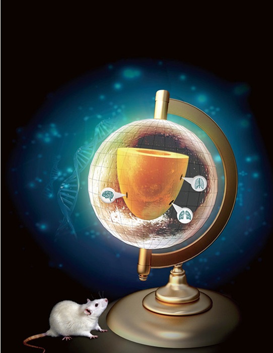
Peng et al. described the spatial transcriptome of the mouse gastrula that, in addition to delineating the gene expression profile of cell populations at defined positions, provides a global positioning system to pinpoint the best-fit address of single epiblast cell of interest in the embryo, thereby revealing its prospective developmental fate and the relevant gene network activity. The latitude and longitude lines in the terrestrial globe indicate the positional information revealed by the spatial transcriptome and the location pointers indicate the three represented developmental fates of the mid-gastrulation mouse embryo. (Image by JING Naihe’s lab)
