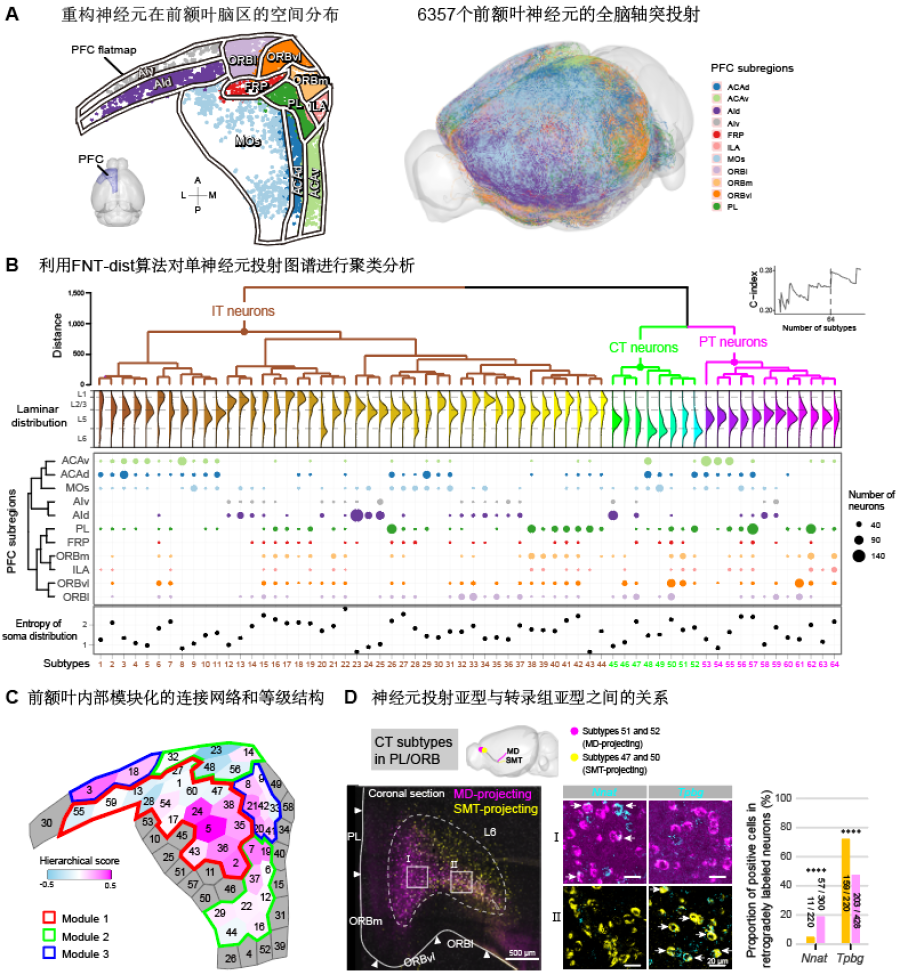
A recent study published in Nature Neuroscience reported the first release of the whole-brain projectome of over 6000 single neurons in mouse prefrontal cortex (PFC), the largest database of whole-brain single-neuron projectome of mouse to date. Through comprehensive analysis, this study identified 64 projectome-defined neuron subtypes in the mouse PFC and their spatial organization, the modularity and hierarchy of intra-PFC connectivity, and the correspondence between transcriptome-defined and projectome-defined neuron subtypes. The study established a comprehensive single-neuron projectome of mouse PFC, systematically studied the internal connectivity and the efferent projection patterns of PFC, and proposed a working model of the PFC, thus providing a structural basis for the neural mechanisms of high-level cognitive functions of the PFC. The study also established the pipeline for future studies of whole-brain mesoscopic projectome in model organisms.
This study was a collaboration between Dr. YAN Jun’s lab and Dr. XU Ninglong’s lab at the Institute of Neuroscience, State Key Laboratory of Neuroscience, Center for Excellence in Brain Science and Intelligence Technology of the Chinese Academy of Sciences, and Dr. GONG Hui’s lab at Wuhan National Laboratory for Optoelectronics, HUST-Suzhou Institute for Brainsmatics, Huazhong University of Science and Technology.
The information flow between different brain regions in the cerebral cortex relies on the long-range axonal projections of neurons. The neurons with different projection patterns are often involved in distinct brain functions. Therefore, studying the projection patterns of individual neurons is crucial for the understanding of the organization and information processing in the brain. The whole-brain projections of neurons in the mouse cortex have been previously studied by using the traditional bulk labeling and tracing method. Cortical projection neurons can be largely divided into intratelencephalic (IT) neurons, pyramidal tract (PT) neurons and corticothalamic (CT) neurons based on their whole-brain projection patterns. However, recent studies have shown that within these traditionally defined neuron classes, more complex neuron subtypes might exist and correspond to even finer functional subdivision.
Furthermore, the connectivity between individual neurons provides the essential information for studying how neural circuit regulates different functions. Therefore, reconstructing the whole-brain projectome at single-neuron level will identify new neuron subtypes and uncover wiring rules of brain network, allowing more systematic understanding of how brain works. Currently, there are intense international efforts to study the whole-brain projectome at single-neuron level.
In the brain, PFC is the center of high-level cognitive functions including decision-making, working memory and attention. The abnormality and malfunction of PFC can cause many neuropsychiatric diseases. The axonal projection of PFC covers nearly all brain areas including the cortex, striatum, thalamus, midbrain, and hindbrain. Previous studies on the whole-brain projection patterns of PFC neurons were based on the bulk labeling of neuronal populations. However, the axonal projection patterns of neurons in PFC at single-cell level remain unclear. Although recent studies suggested the existence of modular and hierarchical structures between different brain regions in the mouse cortex, the modular organization of the connectivity network within the PFC remains to be uncovered.
Reconstructing the whole-brain projectome at the single-cell resolution in mammalian brains is a daunting task and requires continuous tracing of single neurons one by one in the large-scale TB-sized light microscopic imaging data in 3D. The entire tracing process is labor-intensive, extremely complex and time-consuming. To solve this problem, Dr. GOU Lingfeng in Dr. YAN Jun’s lab developed a software package, Fast Neurite Tracer (FNT), for high-throughput single-neuron projectome reconstruction and analysis for TB-sized light microscopic imaging data. FNT facilitated the large-scale studies on single-neuron projectome.
In collaboration with Dr. GONG Hui’s lab at Huazhong University of Science and Technology, together with many labs and facilities at the CAS Center for Excellence in Brain Science and Intelligence Technology, the team successfully reconstructed complete axon morphology of a total of 6,357 single neurons in PFC from the optical imaging data of 161 mouse brains (Figure A).
From the PFC single-neuron projectome, the team identified 64 projectome-defined subtypes after quantifying the similarity of axon morphology of different neurons. The team mapped the spatial distributions of these neuron subtypes in different prefrontal subregions and cortical layers (Figure B). In addition, the team analyzed the intra-PFC connectivity network and constructed a high-resolution intra-PFC network, revealing the modular and hierarchical structure within PFC (Figure C). Finally, the team performed comparative analysis between transcriptome-defined and projectome-defined neuron subtypes. Using the retrograde tracing and single-molecule fluorescence in situ hybridization, they found that each transcriptome-defined subtype corresponds to multiple projectome-defined subtypes.
In summary, the team developed a pipeline for efficient single-neuron tracing in TB-sized image data, to reconstruct the single-neuron projectome of mouse PFC. This study identified 64 projectome-defined neuron subtypes in PFC based on the clustering analysis of neural projection patterns and revealed the modular and hierarchical structure of intra-PFC connections by analyzing the intra-PFC connectivity network, providing crucial insights into the understanding of neural mechanism of PFC. Finally, comparison with transcriptome-defined subtypes demonstrated that one transcriptome-defined subtype can correspond to multiple projectome-defined subtypes, further highlighting the importance of single-neuron projectome for neuron classification.
This work entitled “Single-neuron projectome of mouse prefrontal cortex” was published online as a cover paper in Nature Neuroscience on March 31, 2022. This work was carried out by graduate students, GAO Le , LIU Sang , HU Yachuang, and postdoctoral fellow, GOU Lingfeng, under the supervision of Dr. YAN Jun and Dr. XU Ninglong, with the collaborations with Dr. YAO Haishan, Dr. XU Chun, Dr. LI Chengyu, Dr. SUN Yangang and their labs as well as the mouse brain atlas facility at the CAS Center for Excellence in Brain Science and Intelligence Technology. Numerous volunteers participated in testing FNT, especially the group of students led by XIAN Jing and QIAO Yanqing from Shanghai JiGuang Polytechnic College. Dr. GONG Hui’s lab at Huazhong University of Science and Technology acquired all the whole-brain imaging data in this study. Dr. POO Mu-ming , the scientific director of the CAS Center for Excellence in Brain Science and Intelligence Technology, provided valuable advice and guidance of this project. This work was supported by the Shanghai Municipal Science and Technology Major Project Grant and the grants from Ministry of Science and Technology, Chinese Academy of Sciences, and Natural Science Foundation of China.

Figure legend (A) The distribution of 6,357 single neurons in PFC subregions and their axon morphology. (B) The classification of neuron subtypes based on their axon morphology. The distribution of 64 subtypes in different PFC subregions and cortical laminar. (C) The modular and hierarchical structure of intra-PFC network. (D) The correspondence between projectome-defined subtype and transcriptome-defined subtype (CT subtypes in PL-ORB). (Image by CEBSIT)
Keywords: Prefrontal Cortex, Neuron Reconstruction; Single-neuron Projectome
AUTHOR CONTACT:
YAN Jun
Center for Excellence in Brain Science and Intelligence Technology, Chinese Academy of Sciences, Shanghai, China.
E-mail: junyan@ion.ac.cn

